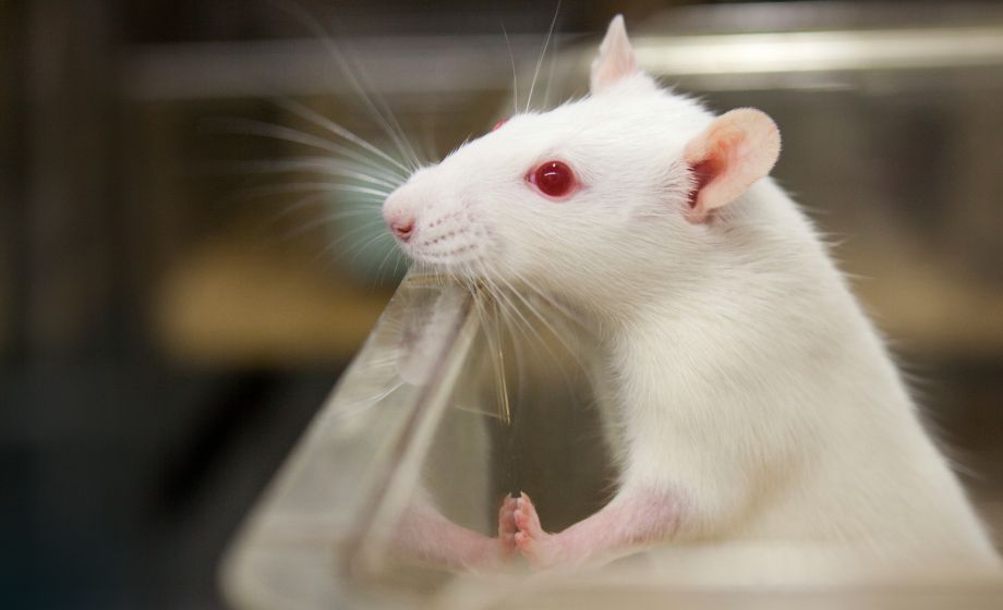
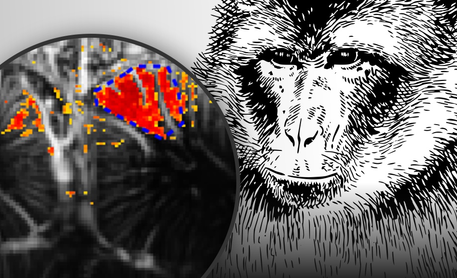
Learn about a unique retinal imaging system optimized for eye research using large animals. Our guest speaker, Dr. David Culp from Powered Research, presents data from his evaluation of aflibercept (Eylea) treatment in a laser-induced model of choroidal neovascularization (CNV) in pigs.
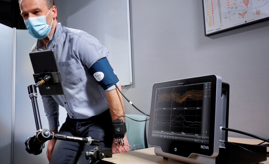
Large animals are an essential research step in the journey from eye drug development to clinical application. The retinas of large animals offer insight into the efficacy and safety of retinal medication. However, using large and low resolution fundus cameras may not reveal the essential details of the treatments' effect. The Micron X large animal camera captures high resolution, detailed images of a range of large animal species, allowing bright field and fluorescein angiography insights into your research.
In this exclusive webinar, sponsored by Phoenix Research Labs, Dr. Christine van Hover introduces the Micron X large animal retinal imaging systems, designed specifically for the challenges of large animal eye and eye-brain research. Dr. van Hover discusses how to take high quality images of the whole large animal retina, what species can be imaged with the Micron X camera and shows examples of fluorescein angiography in non-human primates.
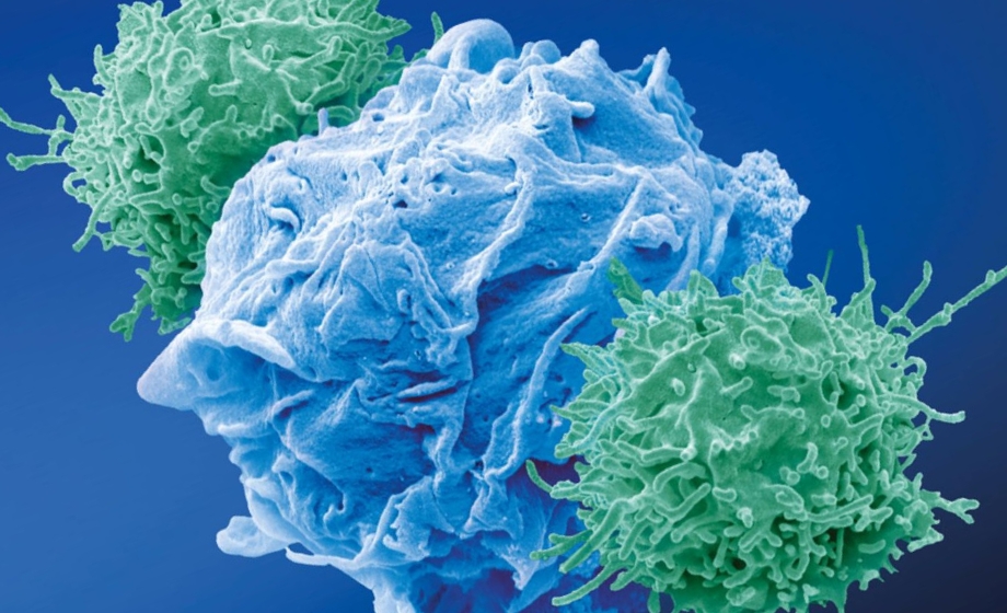
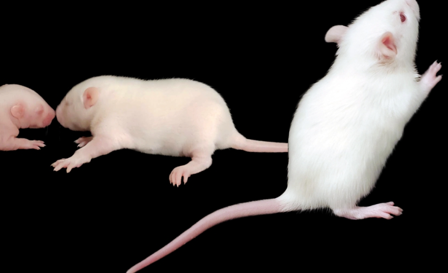
Dr. David Culp, Director of Research at Powered Research, presents data from a recent study designed to evaluate and characterize a porcine model of laser-induced choroidal neovascularization (CNV). CNV complexes were created in weanling pigs, followed by intravitreal injections of aflibercept or balanced salt saline. Dr. Culp provides specific examples of his data where fundus photography, fluorescein angiography (FA), and retinal optical coherence tomography (OCT) were performed. He also shows how the Micron X large animal imaging system produced high resolution fluorescent images to quantify neovascularization. Viewers of this webinar will gain an understanding of how to setup and employ a large animal imaging system in their own laboratory.
Featuring Insights from

David Culp
,
PhD
As the Director of Research for Powered Research, Dr. Culp’s responsibilities include oversight of all scientific programs within the organization. Dr. Culp has a broad background in modeling and analyzing neovascularization, with specific training in analyzing CNV complexes from the Biological Imaging Core Facility in the Division of Intramural Research at the National Eye Institute (NEI).

Christine van Hover
,
PhD
Dr. van Hover earned a PhD in neuroscience at the University of Virginia with graduate work tracing the neuroanatomy of stress and food intake control pathways in the hypothalamus. As Chief Scientist at Phoenix Research Labs, she trains researchers in retinal imaging techniques and provides scientific input into product design.
Sponsored by