Experts discuss how functional ultrasound imaging in awake head-fixed mice can help advance basic neuroscience and neurovascular research, as well as preclinical drug discovery.
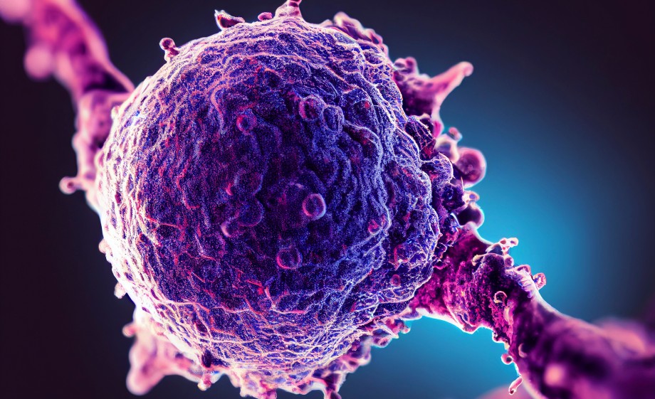
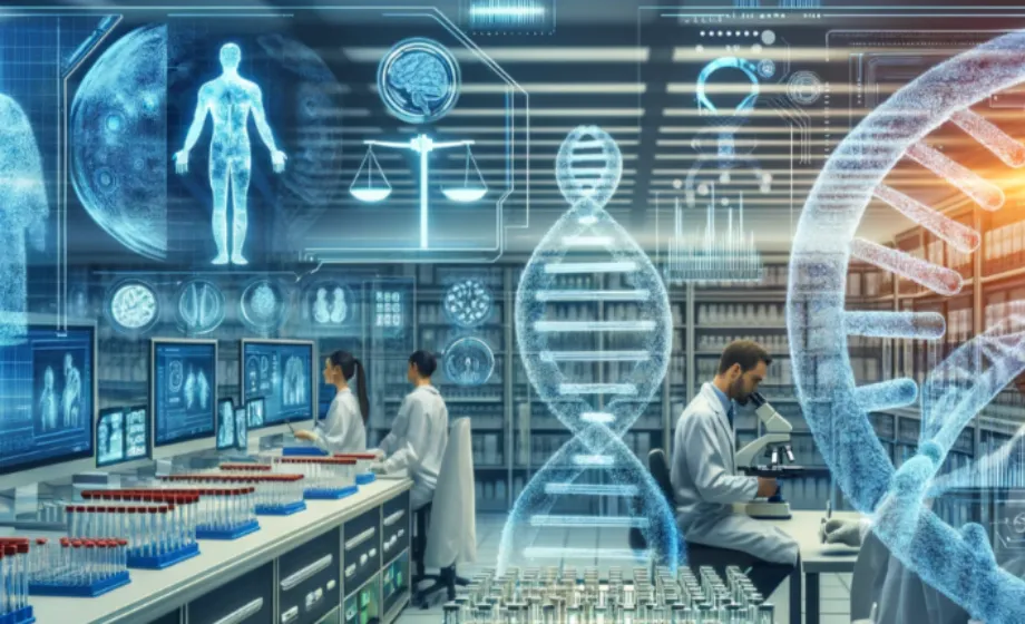
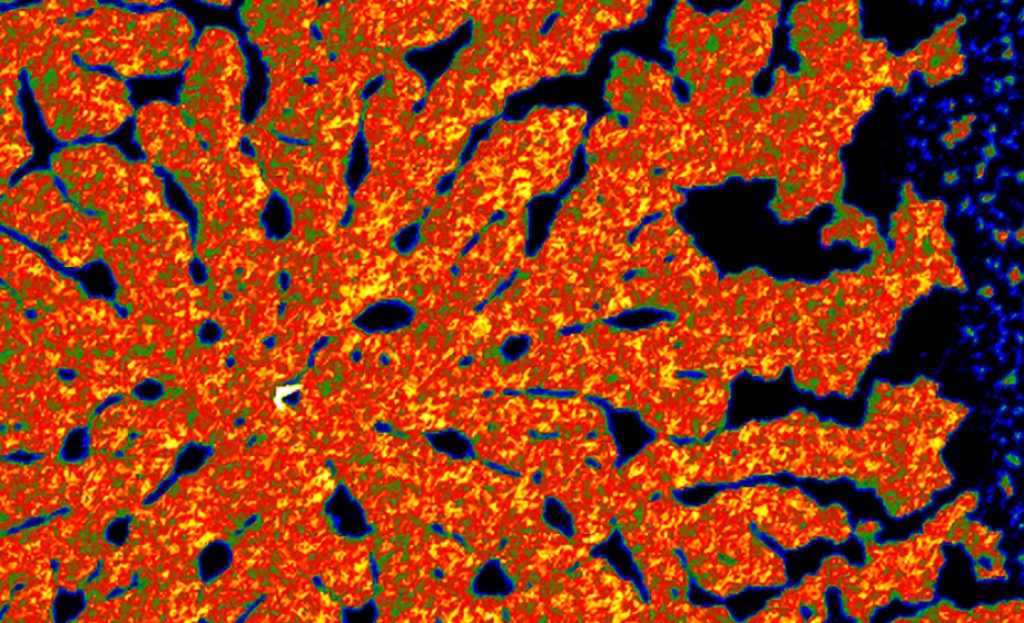
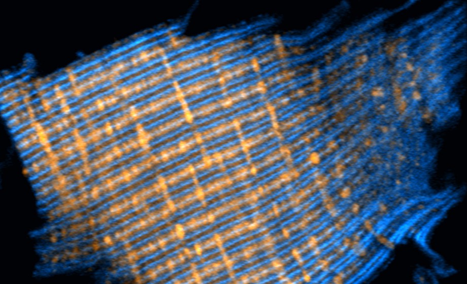
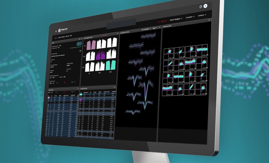
Functional ultrasound (fUS) imaging is a new kid on the block in neuroimaging. It combines high spaciotemporal resolution with deep tissue penetration, which enables non-invasive whole-brain imaging in mice.
This exciting new technology complements and extends classical imaging modalities: it enables more straightforward, unobstructed and non-invasive functional measurements in mouse models of CNS diseases. Sensitive to changes in cerebral blood volume, fUS imaging is used to characterize brain networks with functional connectivity analysis and to measure the responses to sensory stimuli and pharmacological challenges.
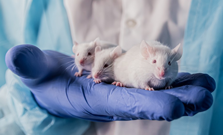
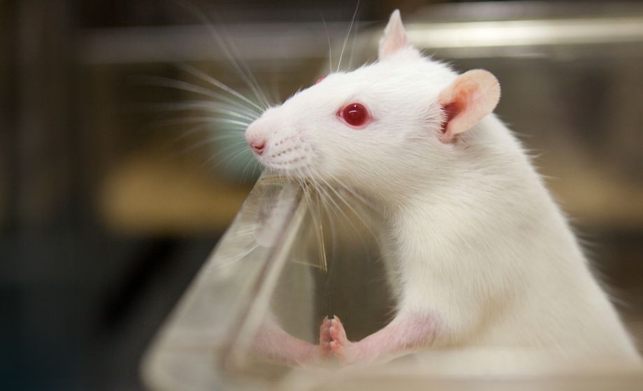
fUS imaging performed in the brain of awake mice removes the biases and artifacts associated with the use of general anesthesia, which is no longer a "necessary evil" of translational imaging. Besides that: fUS imaging in awake mice allows integrating functional imaging with behavioral readouts starting from open field locomotion tracking to maze navigation and sociability studies.
Featuring Insights from

Artem Shatillo
,
MD
Artem Shatillo, MD is a Head of Magnetic Resonance Imaging at Finland site of Discovery from Charles River – one of the world’s leading preclinical CROs. After completing his medical training with major in clinical psychiatry, Artem has switched to experimental neuroscience. With ten years of experience in preclinical imaging, he is now helping our clients to choose and apply the most suitable imaging techniques to approach their questions. His primary research interests include application of novel in-vivo imaging techniques, mainly functional and pharmacological magnetic resonance imaging (f/phMRI) and functional ultrasound (fUS) in animal models of CNS diseases.
Zsolt Lenkei
,
MD, PhD
Zsolt Lenkei, MD PhD is an internationally recognized specialist of brain cannabinoid receptors, as demonstrated by invited lectures world-wide and by his recent election as co-chair of Gordon Conference on Cannabinoids 2019. His team is specialized to investigate the interrelation of neuronal cell biology and neuropharmacology, by using the presynaptic cannabinoid receptor CB1R as a model GPCR. A competitive advantage of the group is the development and use of novel quantitative imaging methods for neuronal cell biology. Since 2012 a strong new line of research was initiated in collaboration with the team 2 for the development of novel ultrafast ultrasound-based in vivo imaging modalities, with results jointly published in Nature Communications, NeuroImage and Nature and co-founding of a start-up company.
Sponsored by
.png)