Scientists present data on 2-photon imaging of hippocampal place cells and on stress monitoring in head-fixed, awake and behaving mice.
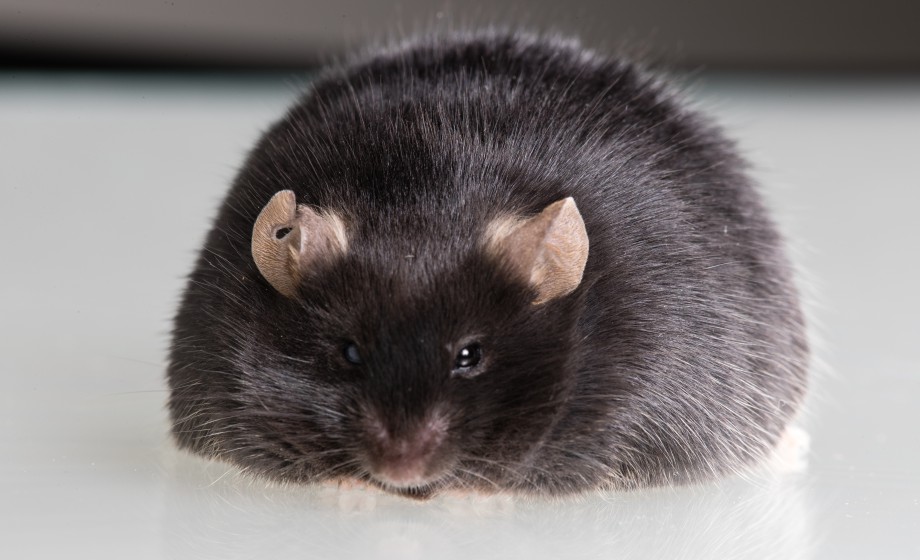
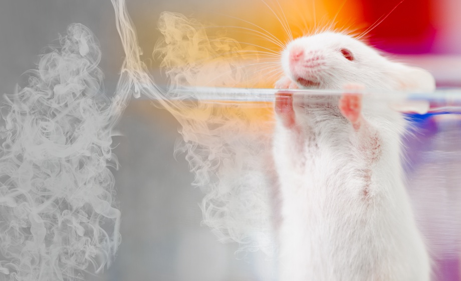
Head fixation is a powerful approach for studying the rodent brain in vivo. Stabilizing the head of a freely moving and behaving animal allows the researchers to study neuronal activity using single-cell electrophysiology and 2-photon imaging. These movement-sensitive techniques are inherently challenging for experiments with behaving rodents, unless they are aided by instrumentation that provides both stability and stress-reduction.
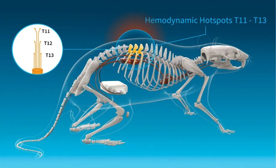
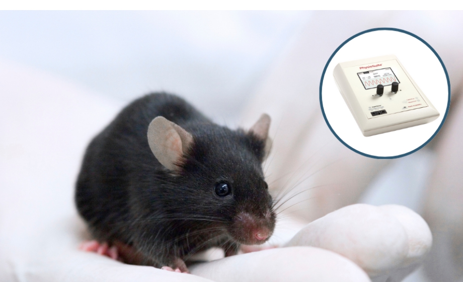
In this webinar sponsored by Neurotar, experts present their research on 2-photon imaging of hippocampal place cells and on stress monitoring in head-fixed awake behaving mice. Dr. Konrad Juczewski from the National Institutes of Health (NIH)/National Institute on Alcohol Abuse and Alcoholism (NIAAA) discusses the impact of head fixation on animal's stress, locomotion and performance in classical behavioral paradigms.
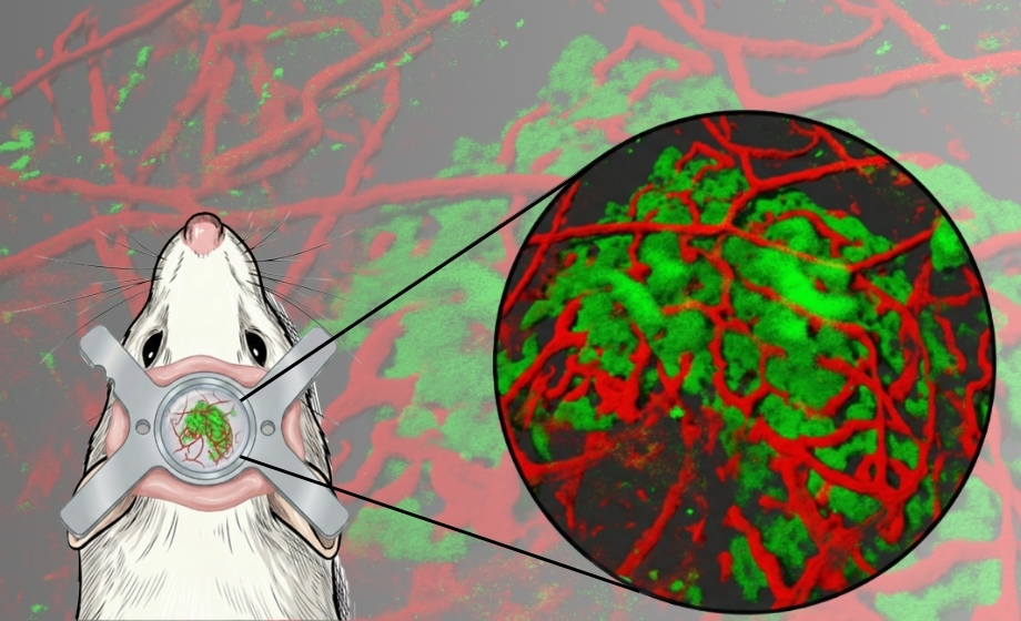
Dr. Mary Ann Go from the Laboratory of Neural Coding and Neurodegenerative Disease at Imperial College London led by Prof. Simon Schultz presents her research using 2-photon microscopy aimed at place cell mapping in the hippocampus during exploration and navigation of a circular linear track.
Featuring Insights from

Konrad Juczewski
,
PhD
Dr. Konrad Juczewski’s research at Prof. David Lovinger’s lab is focused on examining the mechanisms involved in synaptic plasticity and the function and roles of cortico-basal ganglia circuits in habit formation and addiction. In particular, Dr. Juczewski is interested in understanding how sensory processing and plasticity mechanisms are changed by substance abuse and by neurodevelopmental disorders.

Mary Ann Go
,
PhD
A major focus of Dr. Go’s is to better understand how information is processed by mammalian neural circuits underlying memory. In particular, she uses tools to measure neuronal activity while an animal is performing a spatial memory task with the ultimate aim of characterizing mouse models of Alzheimer’s Disease and gaining a better understanding into the effects that proposed therapies have on hippocampal-neocortical circuits.
Sponsored by