Selina Soyeon Ahn, PhD introduces IVIM Technology's All-in-One real-time intravital confocal and two-photon microscopy system and its application to immunology and neuroscience research.
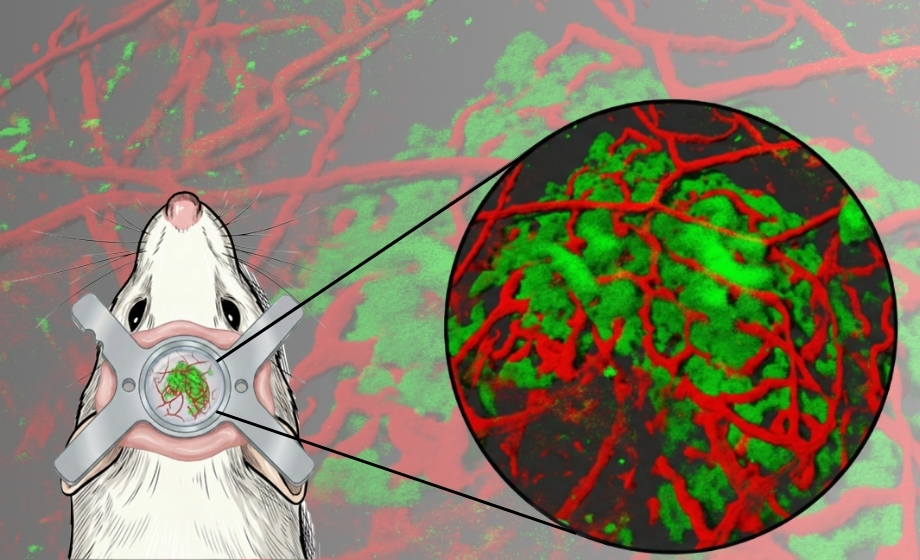
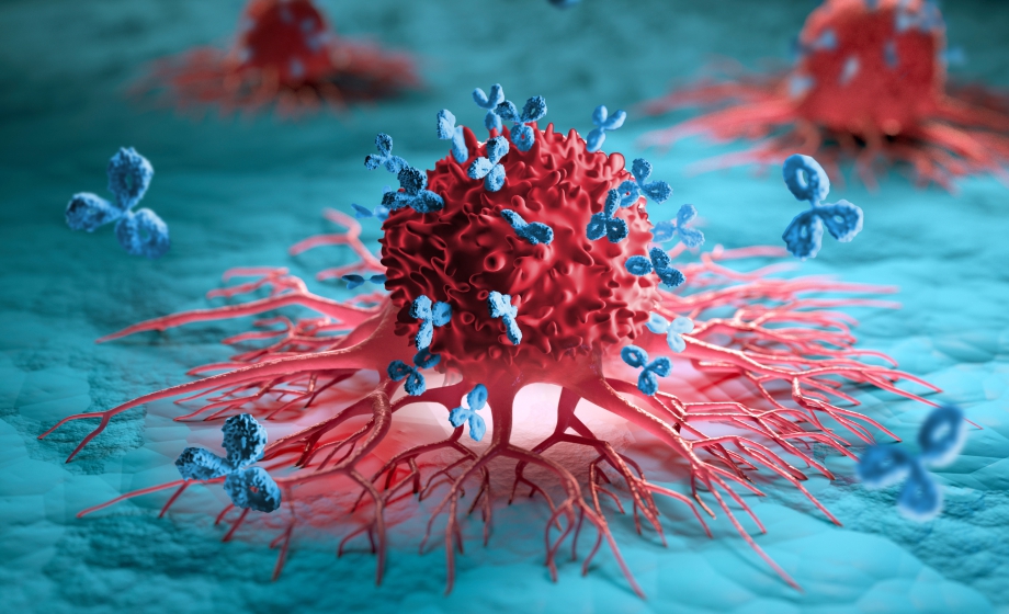
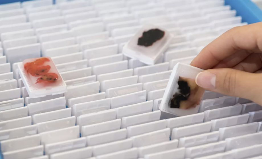
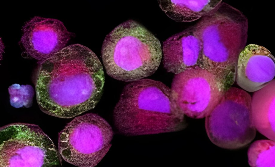
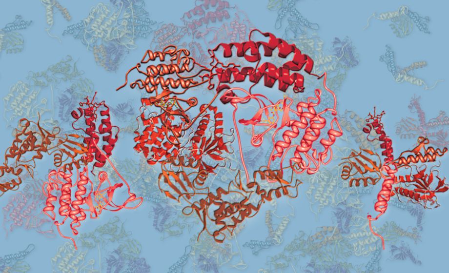
Intravital microscopy is a unique imaging technique to visualize various in vivo cellular-level dynamics such as cell trafficking, cell-to-cell, or cell-to-microenvironment interactions in a live animal. Intravital imaging of cellular dynamics in a natural physiological microenvironment can provide unprecedented insights into the dynamic pathophysiology of human diseases that were impossible to obtain through conventional histological observation of ex vivo samples or in vitro culture systems. It is a unique tool for the development of new therapeutics and diagnostics by providing improved accuracy and reliability for in vivo target validation with delivery monitoring and efficacy assessment. It has been used to directly analyze the delivery and efficacy of new biopharmaceuticals such as antibodies, cell therapy, gene therapy, nucleic acids, and exosomes in an in vivo microenvironment.
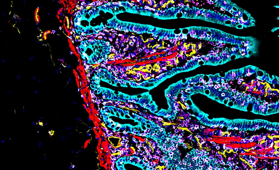
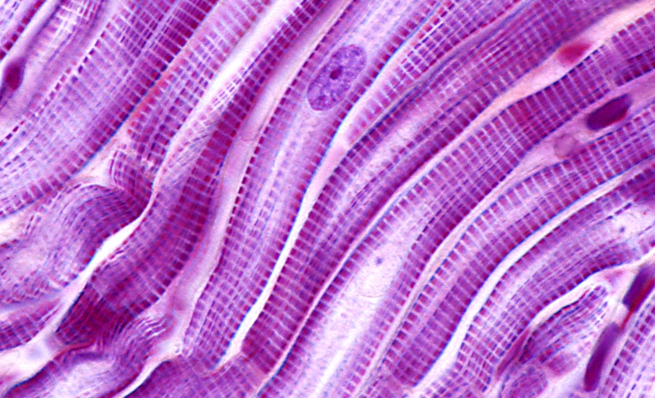
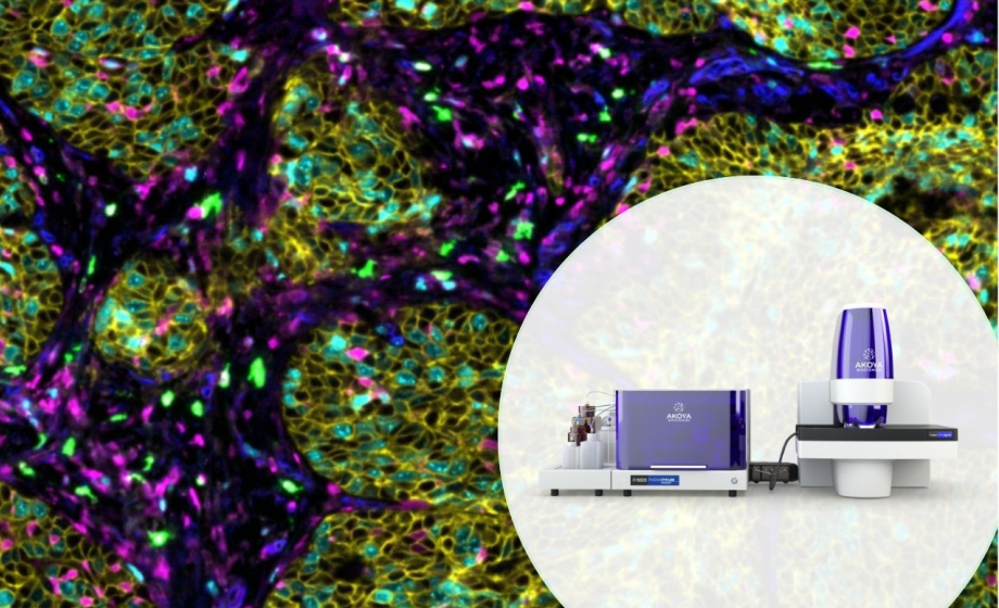
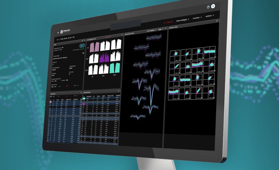
IVIM system has been extensively optimized for in vivo cellular-level imaging of most organs in live animal models for various human diseases, which can acquire a real-time multi-color fluorescence microscopic image in sub-micron resolution with automated motion compensation. Intravital imaging application to various organs is fully available with the IVIM system, in which we can get tons of dynamic information from individual cells in the target tissue/organ in living animal. In this webinar, recent studies are introduced utilizing the real-time intravital imaging technique to investigate dynamic cellular-level pathophysiology of various human diseases.
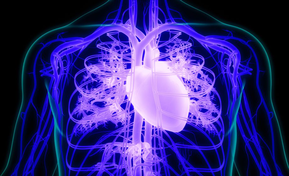
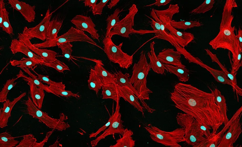
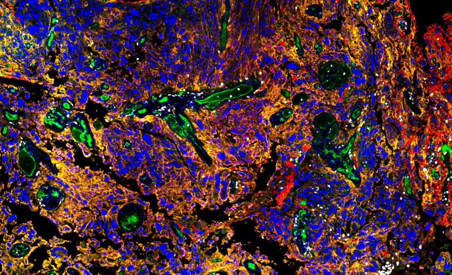
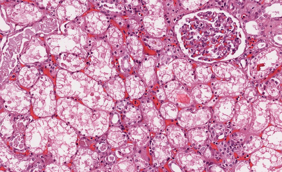
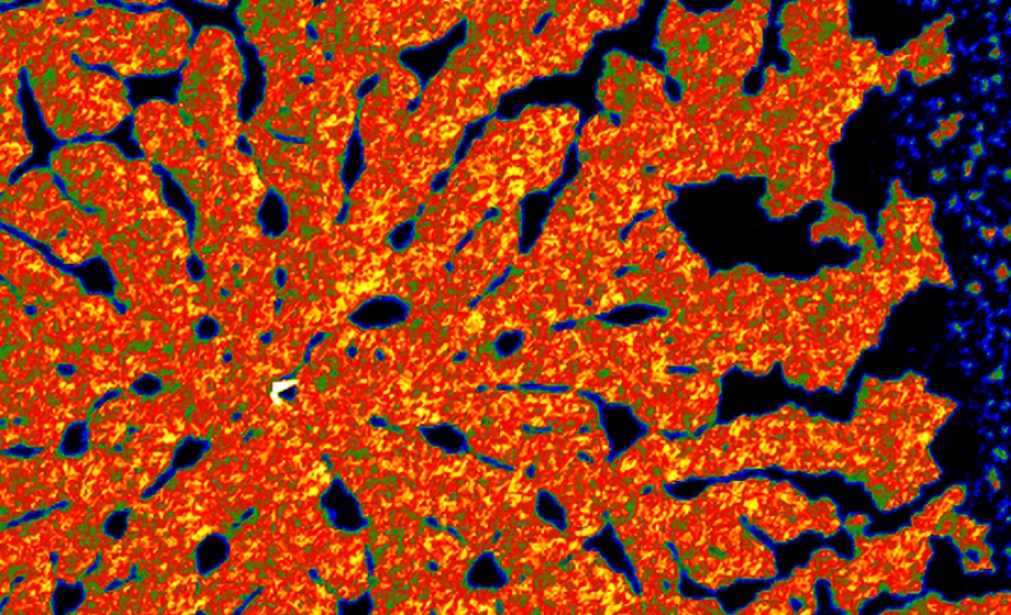
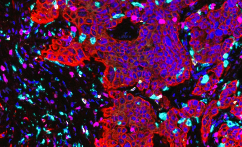
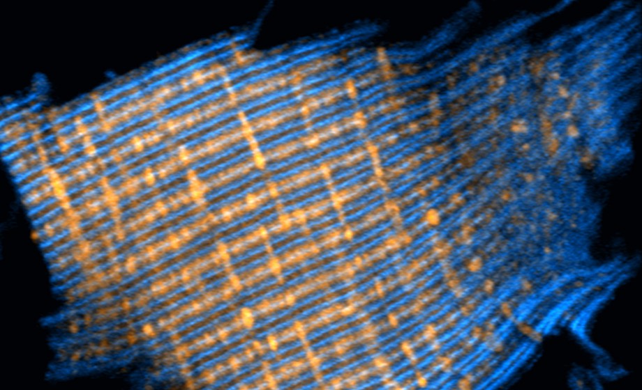
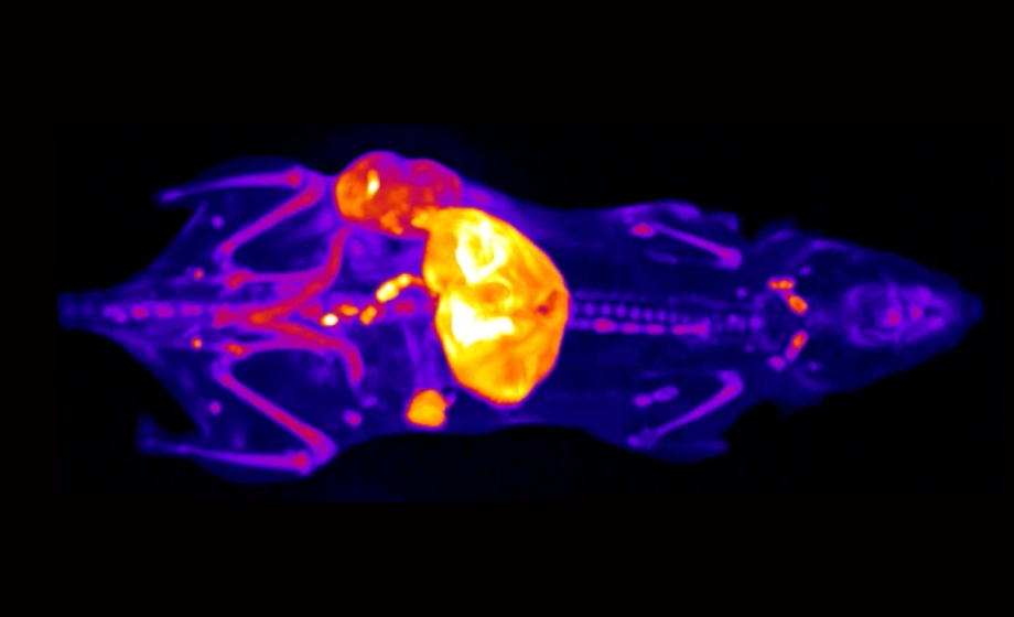
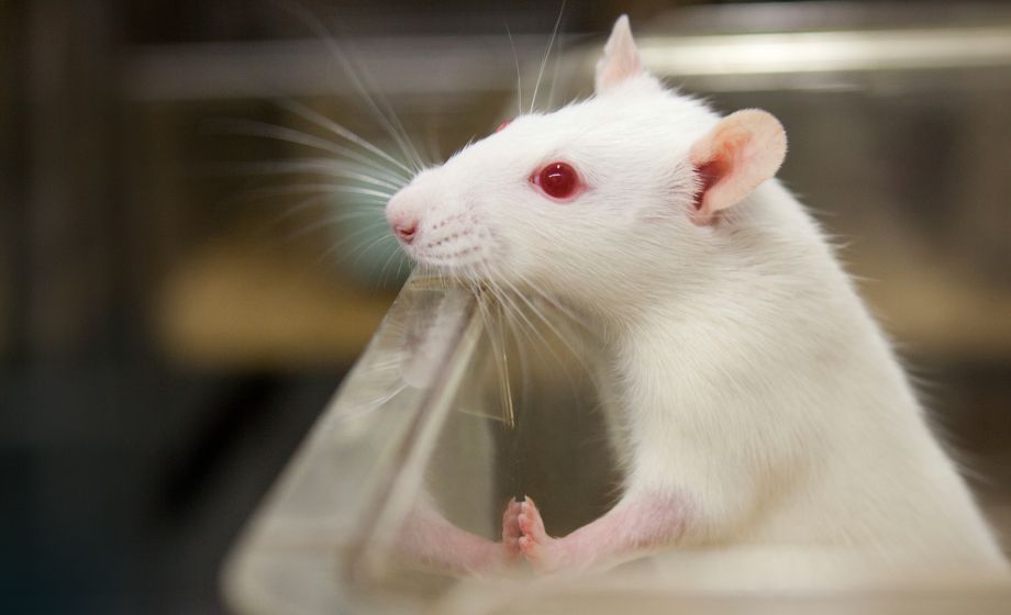
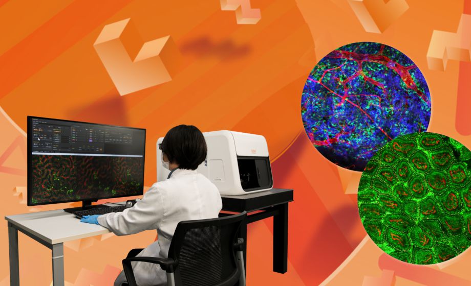
The virtual demo includes the imaging of the liver using specific markers: H2B-GFPxActinDsRed for cellular visualization and CD31-F647/C for vascular visualization. Additionally, we feature brain imaging using the WT CIW model. The fluorescent markers FITC or EB/MS are used for visualizing specific cellular components or structures in the brain.
Featuring Insights from

Selina Soyeon Ahn
,
PhD
Selina Soyeon Ahn, PhD has been working in the intravital imaging research field since 2014. As an application specialist at IVIM Technology, she provides hands-on practical support for scientists in various research fields to take full advantage of intravital imaging techniques to maximize their research performance.
Sponsored by
.png)