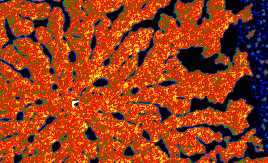
Webinar Summary
- Describe Shear Wave Elastography
- Discuss the value of Elastography in the clinic, and preclinically
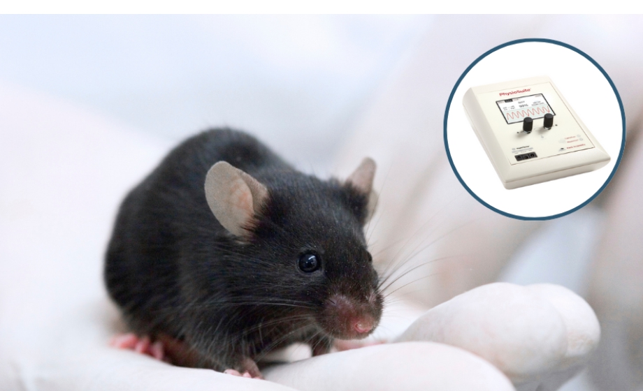
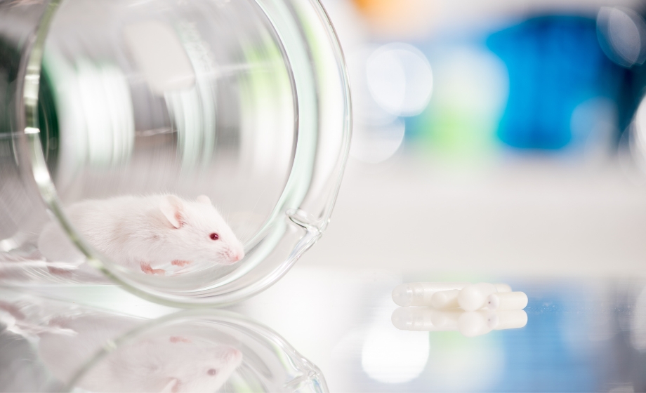
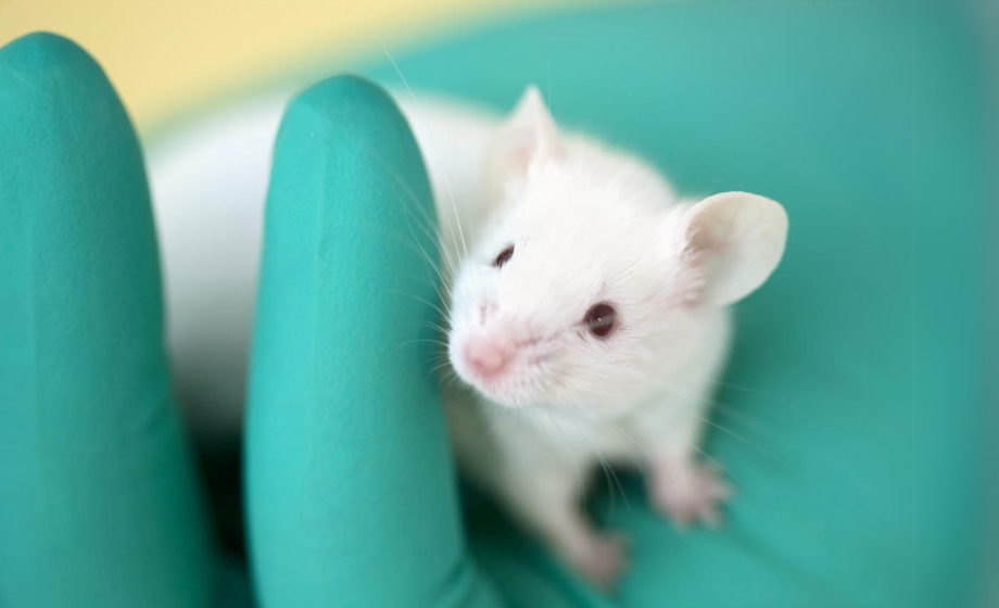
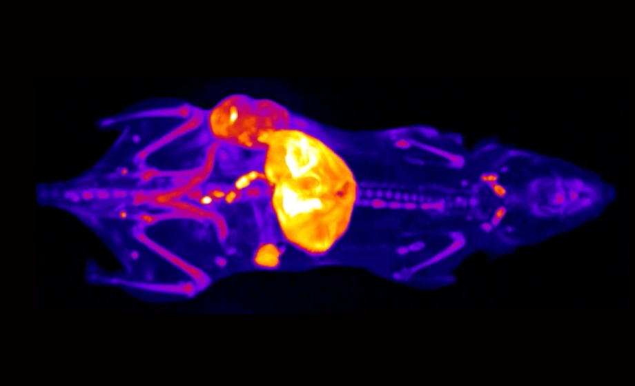
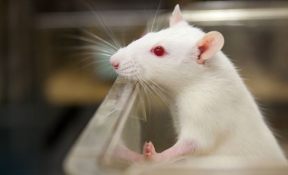
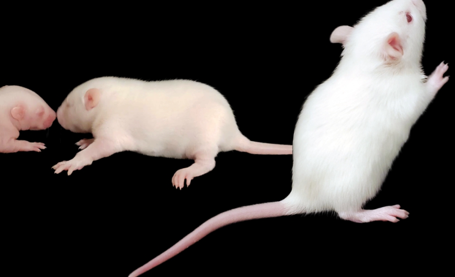
- Showcase Elastography with live imaging of a mouse liver
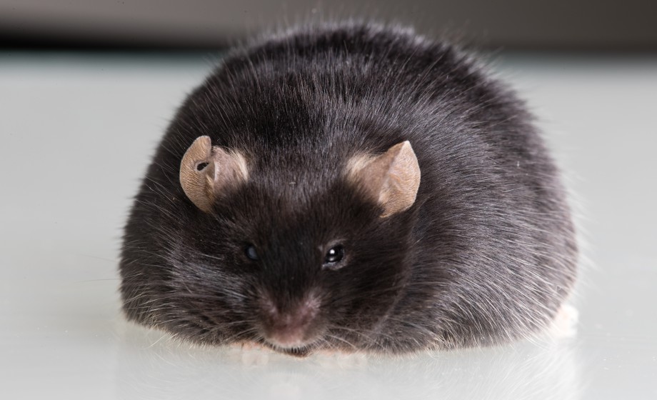
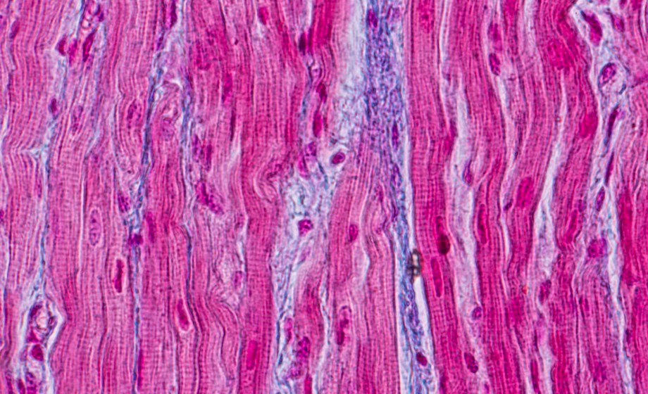
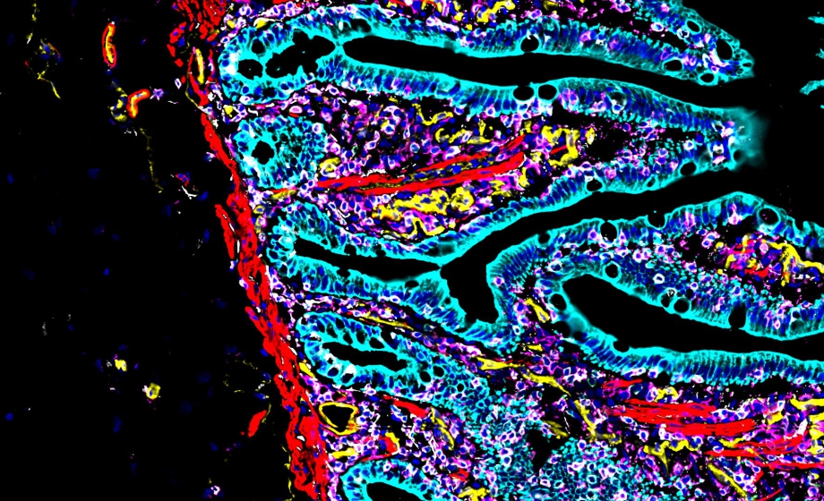
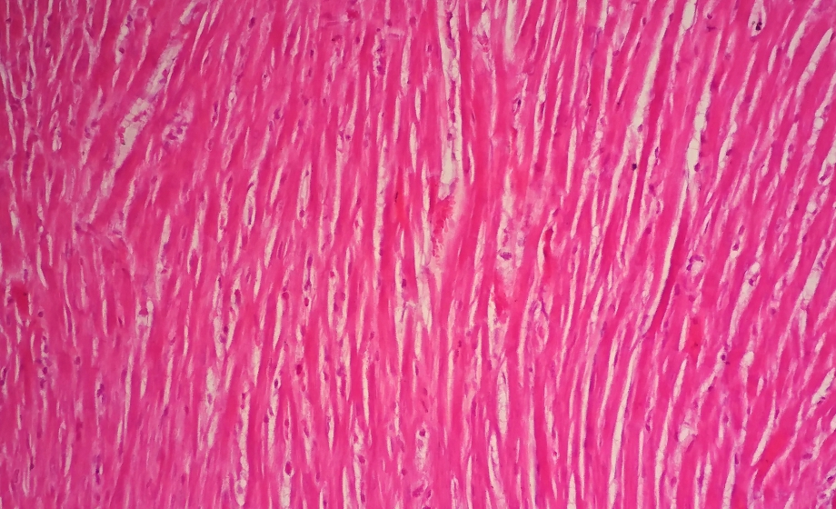
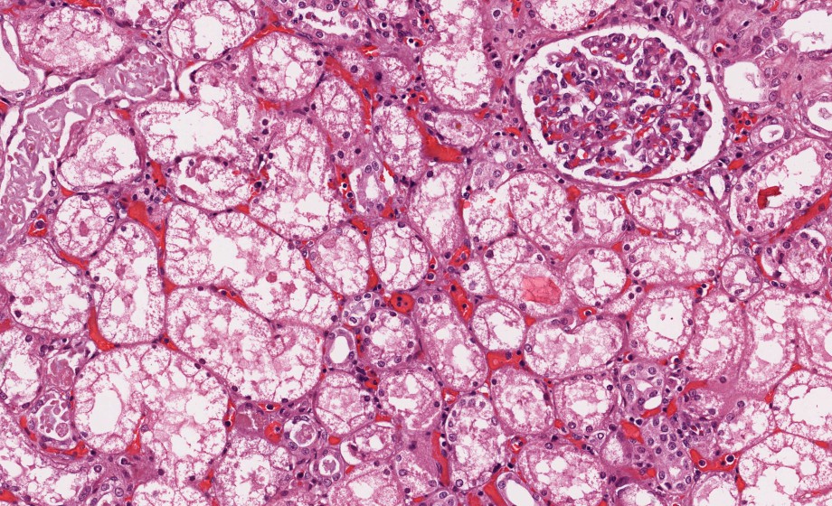
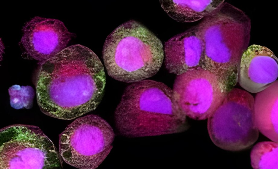
- Share images of normal vs fibrotic livers
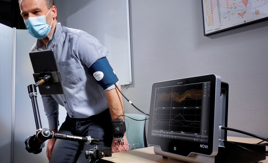
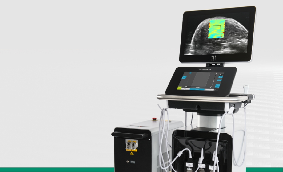
Elastography is an established method for visualizing and assessing liver fibrosis and tissue stiffness in the clinic. This powerful tool is now available at high frequency, specifically optimized for small animal imaging. Following this presentation, there will be a live Q&A Session where audience questions will be addressed.
Featuring Insights from
Kristiina Aasa
,
PhD
Kristiina Aasa completed her M.Sc. in Anatomy and Ph.D. in Biomedical and Molecular Sciences, both at Queen's University. She joined FUJIFILM VisualSonics 10 years ago as an Applications Scientist and is now the Product Manager.
Isabel Newsome
,
PhD
Dr. Isabel (Izzie) Newsome is a Scientific Applications Specialist for Fujifilm VisualSonics. In 2021, she completed her Ph.D. in biomedical engineering at the University of North Carolina at Chapel Hill in the lab of Dr. Paul Dayton, where her dissertation focused on novel contrast-enhanced ultrasound methods and nonlinear microbubble dynamics in the context of tumor-associated angiogenesis.
Sponsored by