Experts provide a tour of academic and industrial use cases in Materials Science, Engineering and Life Science.
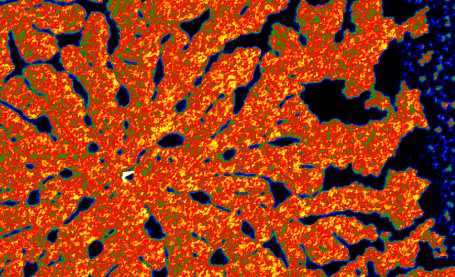
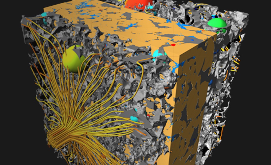
X-ray computed tomography (CT) is becoming an increasingly important tool for the non-destructive characterization and inspection of the three-dimensional microstructure of various materials, products and sample types. The technique creates a three-dimensional representation of a sample/material by reconstructing cross-sectional images or ‘virtual slices' through a sample.
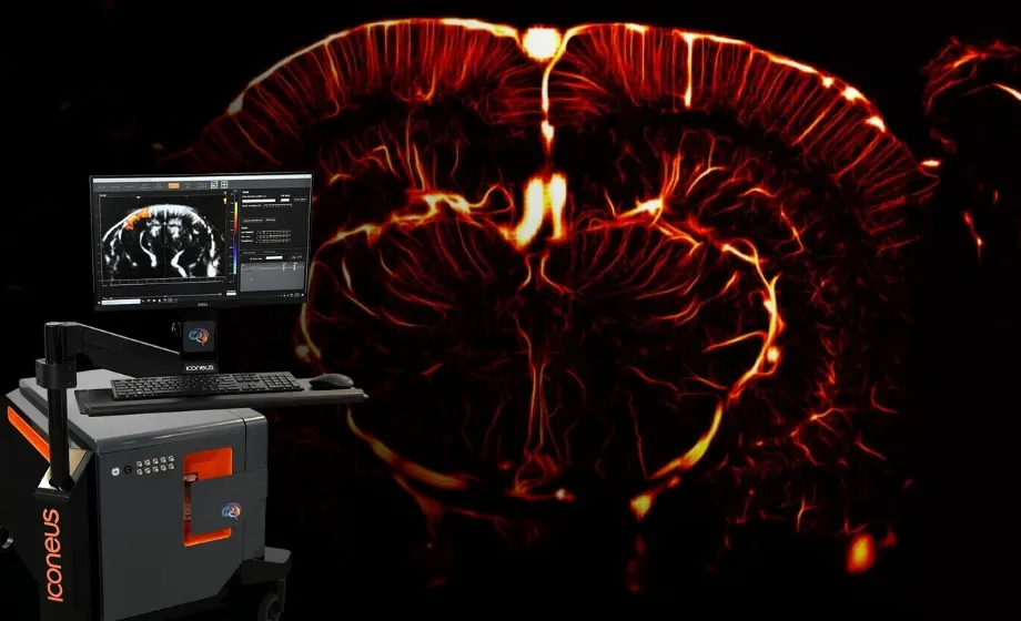
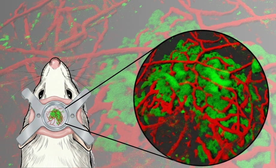
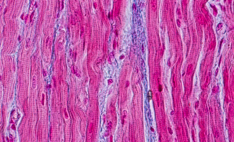
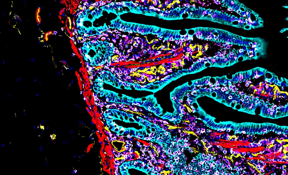
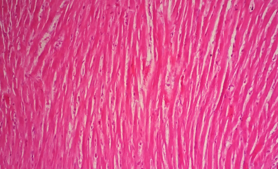
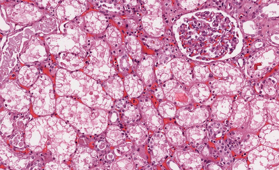
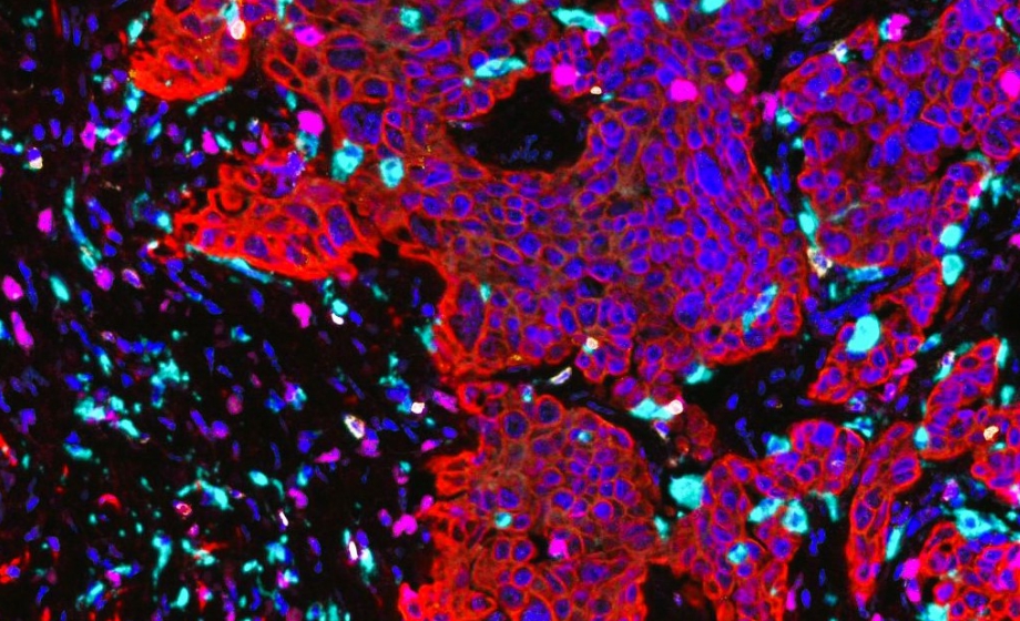
In this webinar, Robert Williams, PhD, and Mark Riccio highlight the versatility of the Thermo Scientific HeliScan microCT, demonstrating the wide breadth of sample types and sizes that the instrument can characterize, such as: polymers, metals, manufactured parts, batteries, rock/porous media, electronics, bone and soft tissue (plants, insects, brain, etc). The HeliScan microCT creates valuable solutions by leveraging a helical scanning technique (found in clinical CT scanners) for large volume data acquisition and features a Lab6 X-ray filament for high resolution (400nm) capability.
The ease of use and high throughput of this system makes it ideal for investigations that need to identify and quantify a sample's 3D internal structure (e.g. voids, cracks, pore networks, coatings, etc.) non-destructively. 4D structural dynamics can be studied by acquiring multiple 3D microCT datasets. Additionally, HeliScan microCT is an integral component of a multi-modal macro-scale to atomic-scale workflow involving focused ion beam/scanning electron microscopes and transmission electron (TEM) microscopes.
Featuring Insights from
Robert E. A. Williams
,
PhD
Robert E. A. Williams is the Assistant Director for Research and Development for the Center of Electron Microscopy and Analysis (CEMAS) at The Ohio State University.
Mark Riccio
Mark Riccio is a Product Marketing Manager for X-ray microCT at Thermo Fisher Scientific’s Materials & Structural Analysis Division.
Sponsored by