Extracellular Vesicles as Breast Cancer Biomarkers: Diagnostic Potential and Recent Mechanistic Findings Blog post by Nina Culum, MSc Breast cancer is recognized as the most prevalent type of cancer in the world, and is the most commonly diagnosed cancer among American women [1, 2]. Triple-negative breast cancer (TNBC), which lacks estrogen, progesterone, and human epidermal growth factor receptor 2 (HER2), is particularly aggressive, challenging to treat, and accounts for 15-20% of all breast cancers [3]. In this blog, we review a study by Xie et al. published in Nature Communications, in which they examined the transforming growth factor-_ (TGF-_) targetable signaling pathway and its role in malignant cancer progression and immune suppression [4].
Tumor-derived extracellular vesicles as non-invasive cancer biomarkers
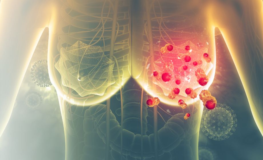
We discussed extracellular vesicles and their applications to vaccine development in a recent blog post , but they are also of interest as cancer biomarkers. Tumor-derived extracellular vesicles (TEVs) carry bioactive cargo such as nucleic acids and proteins from their parent cells during metastasis and mediate intercellular communication, making them potential diagnostic markers for tumor development [5]. Although exosomal TGF-_ released from cancer cells has been shown to favor invasive growth and metastasis, Xie et al. noted that it is unknown whether TEVs containing TGF-_ can influence T cell exhaustion in the tumor microenvironment [4]. The authors also demonstrated that high levels of TGF-_ type II receptors (T_RII) mark circulating extracellular vesicles (crEVs) from malignant breast tumors (Figure 1). https://insidescientific.com/wp-content/uploads/2022/08/TEM-images-of-breast-cancer-EVs-1.png
Figure 1: Transmission electron microscopy images of breast cancer cell line-derived extracellular vesicles immunogold-labeled with T_RII antibodies (scale bars = 50_nm), in which many of the T_RII molecules were found to be anchored to vesicle membranes. 2022, Xie et al. , licensed under CC BY 4.0 .
Diagnostic and prognostic relevance of T_RII+ crEVs in breast cancer
The secretion of T_RII by extracellular vesicles was investigated both in murine models of breast cancer as well as in breast cancer patients. In mice, blood levels of T_RII+ crEV positively correlated with tumor burden, increased proportionally with tumor size, and were associated with metastasis. In the sera of healthy human donors, crEVs exhibited very low baseline T_RII positivity, which was much higher in 89% of breast cancer patients. Additionally, the amount of T_RII in crEVs was the highest in TNBC patients compared to HER2+ and luminal patients. Patients with distant metastasis also exhibited higher amounts of T_RII in crEVs than those without metastasis. The authors determined that the percentage of T_RII+ crEVs could be a classifier for breast cancer, as they were able to differentiate between breast cancer patients and healthy donors with 92% accuracy, 93% sensitivity, and 90% specificity. Furthermore, patients with lower levels of T_RII+ crEVs also exhibited improved overall survival and metastasis-free survival compared to those with higher T_RII+ crEVs levels. Results from these experiments indicate that T_RII is a noninvasive biomarker for metastatic breast cancer with both diagnostic and prognostic potential.
Effects of T_RII on metastasis, anti-tumor immunity, and T cell exhaustion
This paper also reports several mechanistic studies that examine the effects of T_RII+ crEVs on metastatic tumor outgrowth and anti-tumor immunity. The authors determined that T_RII transferred by TEVs induces TGF-_/SMAD activity, and that T_RII+ extracellular vesicles play a critical role in cancer stemness and metastasis. Furthermore, breast cancer cells pre-treated with T_RII+ extracellular vesicles were found to be more resistant to the chemotherapy drugs paclitaxel and doxorubicin. The authors also found an inverse correlation with the amount of T_RII+ crEVs and interferon-_ production, suggesting that T_RII+ secretion from tumor cells may negatively affect anti-tumor immunity. Lastly, the authors investigated the relationship between T_RII and CD8+ T cell exhaustion. T_RII depletion in tumors was associated with higher frequencies of effector cytokines, higher levels of inhibitory receptors, and decreased proliferating capacity of CD8+ T cells, suggesting that T_RII from TEVs may induce T cell exhaustion and suppress anti-tumor immunity. Mechanistically, the authors determined that SMAD3 partners with T cell factor-1 (TCF1) to induce CD8+ T cell exhaustion (Figure 2). [fusion_builder_column type="1_1" layout="1_1" align_self="auto" content_layout="column" align_content="flex-start" valign_content="flex-start" content_wrap="wrap" spacing="" center_content="no" column_tag="div" link="" target="_self" link_description="" min_height="" hide_on_mobile="small

Take the Next Step
Explore Suppliers
Browse trusted partners with relevant expertise
Review Capabilities
Compare services, experience, and past work
Start Your Project
Connect and begin collaborating


.png)