Functional Ultrasound: Pushing the Boundaries of Neuroimaging Blog post by Andrew Davies, PhD Considerable progress has been made in the field of neuroimaging in recent years, with a number of new and improved modalities now available to researchers, such as functional ultrasound imaging (fUS). Although ultrasound has been used clinically for decades, advances in its temporal and spatial resolution have opened the door for its use in a range of new applications, including neuroscience research [1]. For example, a new method of ultrafast ultrasound was described in 2011 [2], enabling the use of ultrasound for functional measurements in the rodent brain, and has been built upon and expanded ever since. In addition, commercially available systems have now been developed with portability in mind and at a lower cost of entry than many more established methods such as magnetic resonance imaging (MRI) and computed tomography (CT). As a result, the number of studies utilizing fUS for neuroimaging is growing rapidly, and here we review a very recent and outstanding example published in Nature Methods in 2022 from Renaudin et al [3].
Cerebral blood flow and neuronal activity
One of the goals of neuroimaging, regardless of method, has been to study cerebral blood flow (CBF) and its response to neuronal activity, collectively referred to as neurovascular coupling (NVC). As CBF has been shown to be regulated locally, and can be measured non-invasively much more easily than individual neuronal activity, changes in CBF can act as a surrogate marker for function. For example, NVC and CBF responses during memory tasks, or differences associated with specific disease states, can provide key data to elucidate underlying mechanisms and pathological processes in addition to the numerous potential diagnostic applications. In such experiments, increases in spatially restricted CBF can be observed in selectively responsive brain regions, a response known as functional hyperemia.
Functional ultrasound and ultrasound localization microscopy
Ultrasound localization microscopy (ULM) provides static images of the vasculature at a microscopic scale using fUS and microbubble (MB) injections as a contrast agent [4]. In theory, with repeated image acquisition, this approach could be combined with experimental paradigms to measure changes in CBF and functional hyperemia. However, the challenge has been to provide whole brain level images (required for contextualizing data) while maintaining sufficient spatial resolution (to resolve microvessels), at a temporal resolution that is meaningful for the study of neuronal activity (on the order of one second or less). In providing context for their recent studies, Renaudin et al. describe the historical limitations of fUS, ULM, and other neuroimaging methods, such as the need to balance increased spatial resolution with prolonged acquisition times, and the depth of penetration. In this paper, the authors introduce new methods to overcome some of these challenges: functional ULM (fULM).
Experimental protocols, equipment, and results
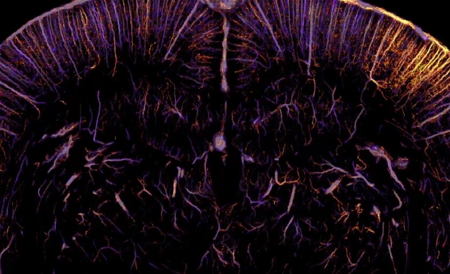
In brief, anesthetized rats were continuously infused with MBs, and responses to mechanical or visual stimuli were recorded transcranially or following craniotomy using an ultrasound probe and scanner (recent webinars demonstrating the applications of similar methods can be found here and here ). The authors provide extensive detail on the critical acquisition and data processing methods, but these are beyond the scope of this review and can be found in the paper. Nevertheless, the data illustrate that these protocols can be used to measure whole rat brain hemodynamic responses to stimuli in vessels as small as 10 m, with only 10 stimuli repetitions, and at a temporal resolution of one second (Figure 1). The results clearly demonstrate that fULM can be used not only as a method to obtain structural images of the microvasculature, but also as a tool to interrogate neuronal activity. Included in the articles supplementary information is a video integrating the data over time, demonstrating the capabilities of the described methods to resolve flux changes in microvessels at the scale of the whole brain in response to stimulation. [fusion_builder_column type="1_2" layout="1_2" align_self="auto" content_layout="column" align_content="flex-start" valign_content="flex-start" content_wrap="wrap" spacing="" center_content="no" column_tag="div" link="" target="_self" link_description="" min_height="" hide_on_mobile="small-visibility,medium-visibility,large-visibility" sticky_display="normal,sticky" class="" id="" background_image_id="" type_medium="" type_small="" order_medium="0" order_small="0" spacing_left_medium="" spacing_right_medium="" spacing_left_small="" spacing_right_small="" spacing_left="" spacing_right="" margin_top_medium="" margin_bottom_medium="" margin_top_small="" margin_bottom_small="" margin_top="0px" margin_bottom="" padding_top_medium="" padding_right_medium="" padding_bottom_medium="" padding_left_medium="" p

Take the Next Step
Explore Suppliers
Browse trusted partners with relevant expertise
Review Capabilities
Compare services, experience, and past work
Start Your Project
Connect and begin collaborating


.png)