Clearing the Brain Fog: Long COVID and Cognitive Impairment Blog post by Andrew Davies, PhD
The COVID-19 pandemic is now approaching its third year, with more than 500 million severe acute respiratory syndrome coronavirus 2 (SARS-CoV-2) cases reported globally. Accompanying the ever-increasing number of COVID survivors is a greater appreciation of the long-term consequences of infection. One frequently reported post-recovery symptom is ongoing cognitive impairment, often referred to as COVID fog or brain fog, which may affect up to 25% of recovered individuals [1]. Although most commonly reported following severe cases, COVID fog is also relatively common among those recovered from mild initial symptoms. In addition to impaired concentration and memory, long COVID has also been associated with increased depression, anxiety, fatigue, and disrupted sleep patterns [2]. Given the global prevalence of SARS-CoV-2 and the detrimental impact of such neurological and cognitive impairment, the public health implications are widespread and considerable. In a recent paper published in Cell, Fernndez-Castaeda et al. examined the underlying neurological changes associated with COVID fog (Figure 1) [3], which we briefly touched on in a recent blog post , and review more extensively here.
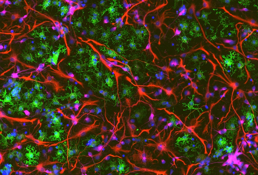
Figure 1: Schematic illustration of the neurobiological effects of respiratory SARS-CoV-2 infection. 2022 Fernndez-Castaeda et al. , licensed under CC BY 4.0 .
COVID fog and chemo fog: a possible clue?
As noted by the authors, the syndrome of cognitive symptoms observed in long COVID bears some resemblance to that observed in patients undergoing certain cancer therapies, leading to their hypothesis that the underlying pathology may also be similar. Notably, microglial reactivity may be affected by some chemotherapy treatments and radiation, resulting in elevated central nervous system cytokine levels, impaired neuroplasticity and neurogenesis, and inflammatory neurotoxicity. They therefore sought to determine whether similar changes in microglial activity and cytokine levels were observed following SARS-CoV-2 infection.
Neuroinflammation and SARS-CoV-2
The authors report that SARS-CoV-2 infection resulted in an inflammatory response in excess of that typically seen with other common viral infections of the respiratory system (e.g., H1N1 influenza). The observed changes in microglial reactivity persisted following COVID, particularly in white matter regions (Figure 2), and elevated levels of the chemokine CCL11 were reported in both animal models and human subjects experiencing COVID fog. Fernndez-Castaeda et al. suggest that the observed selective susceptibility of certain brain regions (e.g., the hippocampus) to elevated levels of CCL11 may at least partly account for differences in the neurological impact of certain viral infections. Loss of oligodendrocytes and myelin were also evident in mouse models of SARS-CoV-2, and the authors note similarities between this pattern and that previously reported in human patients treated with methotrexate chemotherapy.

Take the Next Step
Explore Suppliers
Browse trusted partners with relevant expertise
Review Capabilities
Compare services, experience, and past work
Start Your Project
Connect and begin collaborating


.png)